Beyond 20/20: Eye Doctor in San Juan Capistrano, CA
An eye doctor in San Juan Capistrano, CA can evaluate more than the smallest letters a patient can read by examining eye health, focusing, coordination,…

Glaucoma eye exam in Vienna, VA uses multiple measurements and imaging technologies to look for optic nerve damage that may develop long before a patient notices changes in vision.
This makes glaucoma care a powerful example of how innovation is changing preventive healthcare. Instead of relying on symptoms or a single eye-pressure reading, optometrists can combine structural imaging, functional testing, clinical examination, and data collected over time.
The goal is not merely to generate more test results. It is to identify meaningful changes earlier, determine whether treatment is working, and preserve as much vision as possible.
Glaucoma is a group of eye diseases that damage the optic nerve, which carries visual information from the eye to the brain. The most common form usually progresses gradually and may affect peripheral vision before central vision.
Because one eye may compensate for the other, early changes can be difficult to notice. A person may continue reading, driving, and working without realizing that part of the visual field has been lost.
The National Eye Institute’s glaucoma overview explains that early symptoms are often absent and that a comprehensive dilated eye examination is the only way to determine whether someone has the disease.
Vision already lost to glaucoma generally cannot be restored. Early detection matters because treatment can often slow or prevent additional damage.
Many people associate glaucoma testing with the brief puff of air used to estimate intraocular pressure. Eye pressure is an important measurement, but it cannot diagnose or exclude glaucoma by itself.
Some patients have elevated pressure without detectable optic nerve damage. Others develop glaucoma even though their pressure measurements fall within a statistically normal range.
Pressure can also vary throughout the day and may be influenced by corneal thickness, medications, body position, previous eye procedures, and measurement technique. For this reason, clinicians interpret pressure alongside information about the optic nerve, visual field, drainage angle, and other risk factors.
A glaucoma assessment may include several complementary tests. Each answers a different question, and the combined information creates a more reliable picture of the patient’s eye health.
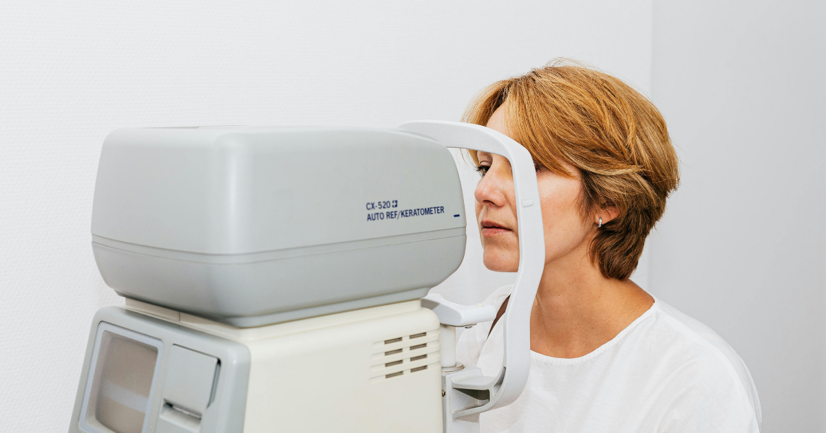
Tonometry measures pressure inside the eye. Different instruments use different methods, including a gentle puff of air or a calibrated probe that briefly contacts the numbed eye.
A single reading provides a snapshot. Repeated measurements help the eye doctor understand whether pressure is stable, fluctuating, or responding to treatment.
Dilating drops enlarge the pupils so the doctor can examine the retina and optic nerve more thoroughly. Glaucoma can produce characteristic changes in the optic nerve’s appearance, but those findings must be interpreted in relation to the individual patient.
Optic nerves naturally vary in size and shape. A larger cup within the nerve does not automatically establish glaucoma, which is another reason longitudinal records are valuable.
Optical coherence tomography, commonly called OCT, uses reflected light to create detailed cross-sectional images of retinal and optic nerve structures. It can measure layers of nerve tissue that may become thinner as glaucoma progresses.
OCT is fast and noninvasive, but the resulting scan is not a stand-alone diagnosis. Image quality, anatomy, severe nearsightedness, and other eye conditions can affect interpretation. The doctor must compare the scan with the examination and other test results.
A visual field test evaluates how well a patient detects lights in different areas while looking at a central target. This provides functional information about peripheral vision.
The test requires concentration and can be affected by fatigue, distraction, blinking, or unfamiliarity. A questionable result may need to be repeated before the doctor determines that a true change has occurred.
Pachymetry measures the thickness of the cornea. This information can help the clinician interpret eye-pressure readings because corneal properties can influence the measured result.
Gonioscopy allows the doctor to evaluate the area where fluid drains from the eye. This helps distinguish open-angle glaucoma from angle-closure conditions and can influence management decisions.
The most valuable feature of modern glaucoma technology is its ability to compare results across time. One OCT scan or visual field test establishes only a momentary baseline. A series can reveal whether the optic nerve appears stable or is changing.
This turns glaucoma management into an ongoing information system:
Computerized comparison tools can flag possible change, but the clinician decides whether that change is real, clinically important, or caused by testing variability. Technology supports professional judgment; it does not replace it.
Anyone can develop glaucoma, but some people require closer attention. Risk factors may include:
The Centers for Disease Control and Prevention identifies family history, age, and certain racial or ethnic backgrounds among important glaucoma risk factors.
Risk does not mean that a person has the disease. It helps determine when testing should begin and how frequently the eyes should be evaluated.
Glaucoma treatment generally aims to lower eye pressure, even when the pressure was not unusually high when the disease was discovered. The appropriate target is individualized according to the type and severity of glaucoma, current pressure, rate of change, age, and other health considerations.
Treatment options may include:
An optometrist may monitor the disease, prescribe appropriate medication within the provider’s scope of practice, and coordinate care with an ophthalmologist when laser or surgical treatment is required.
The strongest plan is not necessarily the most aggressive one. It is the option that achieves sufficient pressure control while considering safety, adherence, cost, side effects, and the patient’s ability to use the treatment consistently.
Eye drops only work when they are used as prescribed. Missed doses, incorrect technique, cost, bottle design, and side effects can all interfere with treatment.
Helpful habits include:
Patients should bring their eye-drop bottles to follow-up appointments when possible. The care team can confirm the product, dosage, schedule, and application technique.
Glaucoma monitoring is customized. A person with stable findings and low risk may be examined less often than someone who recently started treatment or has evidence of progression.
The doctor may schedule visits based on:
Keeping appointments is essential even when vision seems unchanged. Glaucoma can progress without producing obvious day-to-day symptoms.
Patients who move or transfer care should request copies of previous test results, especially optic nerve photographs, OCT scans, visual fields, pressure records, and treatment history.
Historical information gives the new doctor a better basis for comparison. Without earlier records, it may take additional time to determine whether an unusual finding is longstanding or progressing.
A personal record should include medication names, previous laser or surgical procedures, allergies, and the contact information of other eye specialists involved in care.
Most open-angle glaucoma develops gradually, but angle-closure glaucoma can present as an emergency. Sudden severe eye pain, red eye, blurred vision, halos around lights, headache, nausea, or vomiting requires immediate medical attention.
Patients should not wait for a routine appointment when these symptoms appear. Prompt treatment may be necessary to protect vision.
Vienna Eyecare Center is located at 120 Beulah Road NE, Suite 101, in Vienna, Virginia 22180. The practice serves Vienna and nearby communities including McLean, Fairfax, Tysons Corner, and Reston.
Its optometrists provide comprehensive eye examinations and use diagnostic technology to measure eye pressure, evaluate the optic nerve, and monitor changes over time. The practice also provides ongoing glaucoma management and coordinates additional treatment when laser or surgical care is appropriate.
Patients should bring a complete medication list, previous eye records, and information about any family history of glaucoma to their appointment. These details can help establish a more accurate risk profile.
Glaucoma care demonstrates that healthcare innovation is not only about developing new devices. It is also about connecting multiple measurements into a coherent record that supports earlier and more confident decisions.
Eye-pressure testing, optic nerve imaging, visual field analysis, and repeated examinations each contribute different information. When these tools are interpreted together, clinicians can distinguish stable findings from meaningful change and adjust treatment accordingly.
For patients, the most important step remains straightforward: obtain comprehensive eye examinations before symptoms appear and keep every recommended follow-up. The technology can identify change, but timely care allows that information to protect vision.